Facilities
Imaging Facility – versatile imaging with high spatio-temporal resolution
The Imaging Facility enables high-precision exploration of cellular and physiological processes using advanced instrumentation. The laboratory is equipped with two widefield Olympus fluorescence microscopes (IX-83 and IX-73), optimized for functional measurements such as intracellular pH, calcium, sodium and chloride and cAMP level. The IX-83 microscope is equipped with a motorized stage and focus adjustment, as well as a stage-top Ibidi incubator, enabling long-term live cell observations with environmental control. These systems allow real-time visualization of key physiological parameters, offering valuable insights into cellular function and signaling. The facility houses a Zeiss Axio Imager 2 is a high-performance fluorescence microscope designed for demanding biomedical research applications. It delivers exceptional image quality with advanced optics, precise illumination, and reliable automation. This system supports a wide range of fluorescence techniques, enabling detailed visualization of cellular structures and molecular interactions. The system is also equipped with a color camera for the The system is also equipped with a color camera for
the high-resolution digitalization of histology sections and
immunohistochemistry stainings.
The Imaging Facility enables high-precision exploration of cellular and physiological processes using advanced instrumentation. The laboratory is equipped with two widefield Olympus fluorescence microscopes (IX-83 and IX-73), optimized for functional measurements such as intracellular pH, calcium, sodium and chloride and cAMP level. The IX-83 microscope is equipped with a motorized stage and focus adjustment, as well as a stage-top Ibidi incubator, enabling long-term live cell observations with environmental control. These systems allow real-time visualization of key physiological parameters, offering valuable insights into cellular function and signaling. The facility houses a Zeiss Axio Imager 2 is a high-performance fluorescence microscope designed for demanding biomedical research applications. It delivers exceptional image quality with advanced optics, precise illumination, and reliable automation. This system supports a wide range of fluorescence techniques, enabling detailed visualization of cellular structures and molecular interactions. The system is also equipped with a color camera for the The system is also equipped with a color camera for the high-resolution digitalization of histology sections and immunohistochemistry stainings.
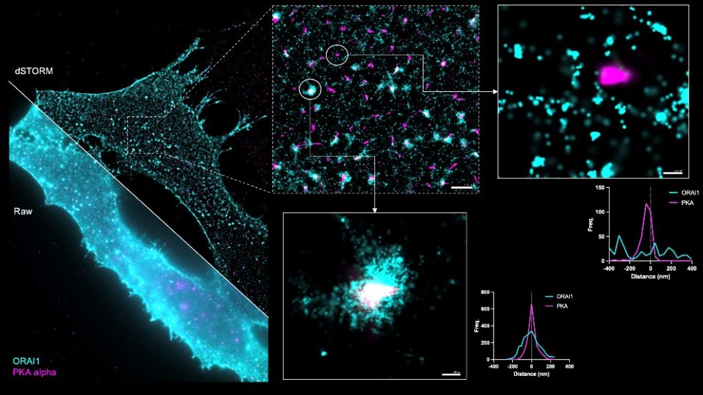
The facility also includes a super resolution microscope from Oxford Nanoimaging. ONI’s Nanoimager is a compact, benchtop super-resolution microscope for single-molecule fluorescence that fits on a desktop and doesn’t require an optical table or dark room. It supports modalities such as dSTORM, PALM, DNA-PAINT, single-particle tracking, TIRF/HILO and smFRET, for both fixed and live-cell experiments. Typical achievable resolution is about 20 nm, enabling quantitative mapping of molecular organization and dynamics. NimOS software and built-in workflows streamline setup and provide real-time data build-up and rapid analyses (e.g., smFRET), making super-resolution accessible on the benchtop.
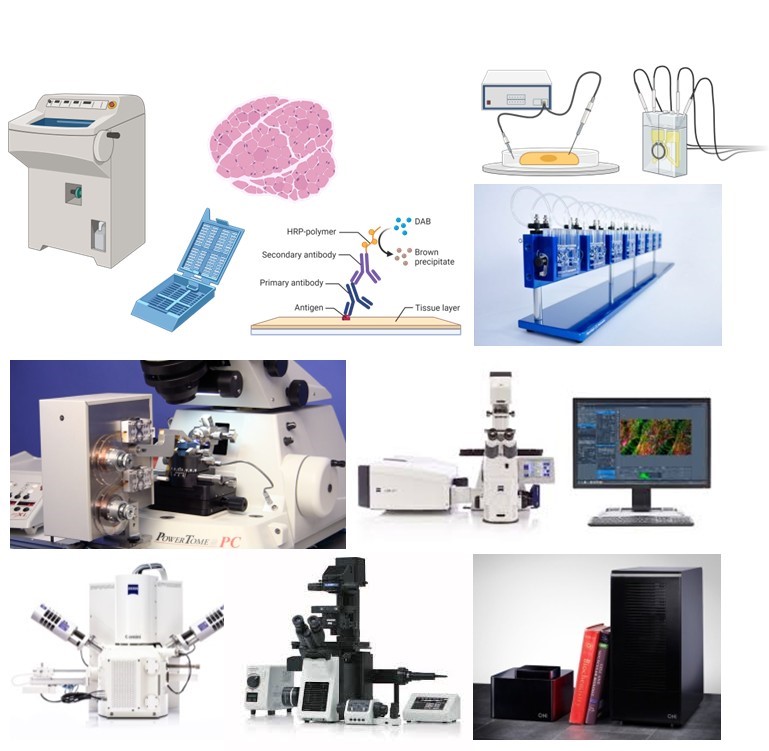
The facility is also equipped with a Ussing chamber system, which is a specialized system for high-precision electrophysiological measurements across epithelial tissues. It enables researchers to investigate ion transport, barrier properties, and transepithelial voltage and resistance under controlled conditions. By providing real-time functional data, the Ussing chamber is an essential tool for studying epithelial physiology, gastrointestinal research, and the mechanisms underlying transport-related disorders.
Together, these instruments create a powerful platform for investigating ion transport, epithelial barrier function, and a wide range of physiological mechanisms.
Animal Facility - Innovative in vivo disease models with reliable results
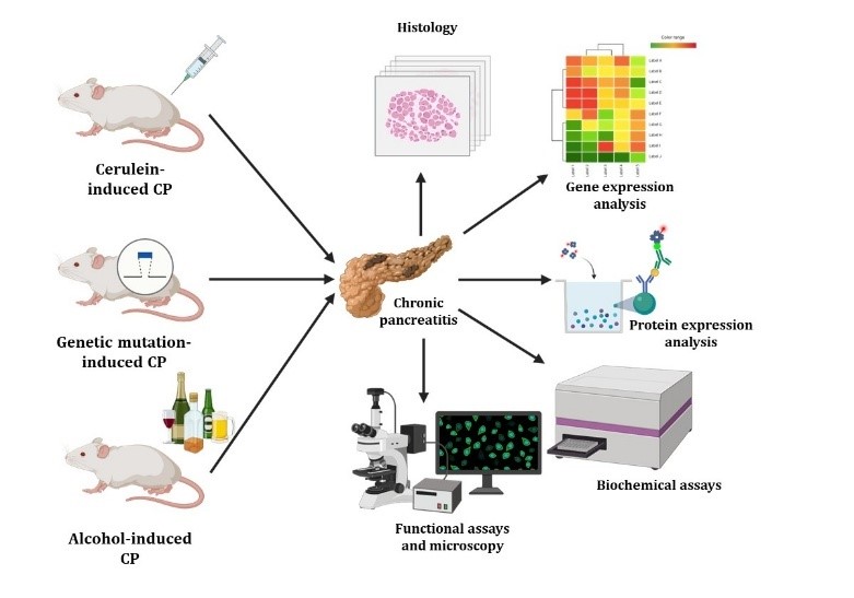
Our state-of-the-art Animal Facility provides a comprehensive and flexible platform for cutting-edge biomedical research and the development of preclinical disease models. A particular focus is placed on models of gastrointestinal disorders, including cerulein-, bile acid- and alcohol-induced acute pancreatitis, cerulein- and alcohol-induced chronic pancreatitis, DSS-induced acute and chronic inflammatory bowel disease, as well as alcoholic hepatitis models. These established in vivo systems enable detailed investigation of disease mechanisms and the testing of novel therapeutic strategies.
The facility houses a wide range of genetically modified mouse strains, from general knock-out strains (such as CFTR KO mice) to advanced conditional knock-out models that allow for tissue-specific modulation of target proteins. These resources support in-depth analysis of physiological processes and pathological alterations, thereby advancing translational research. To ensure the highest standards of experimental work, the facility is equipped with a modern anesthesia machine and a fully operational surgical suite designed for microsurgical procedures. These capabilities guarantee safe, precise, and reproducible interventions in full compliance with international animal welfare regulations and ethical guidelines. In addition, the Animal Facility offers controlled housing conditions
conditions with continuous health monitoring,
specialized technical support from experienced staff, and a collaborative environment that fosters innovation and scientific excellence. Together, these resources make the facility an essential hub for researchers aiming to translate fundamental discoveries into clinically relevant outcomes.
The facility houses a wide range of genetically modified mouse strains, from general knock-out strains (such as CFTR KO mice) to advanced conditional knock-out models that allow for tissue-specific modulation of target proteins. These resources support in-depth analysis of physiological processes and pathological alterations, thereby advancing translational research. To ensure the highest standards of experimental work, the facility is equipped with a modern anesthesia machine and a fully operational surgical suite designed for microsurgical procedures. These capabilities guarantee safe, precise, and reproducible interventions in full compliance with international animal welfare regulations and ethical guidelines. In addition, the Animal Facility offers controlled housing conditions
The facility houses a wide range of genetically modified mouse strains, from general knock-out strains (such as CFTR KO mice) to advanced conditional knock-out models that allow for tissue-specific modulation of target proteins. These resources support in-depth analysis of physiological processes and pathological alterations, thereby advancing translational research. To ensure the highest standards of experimental work, the facility is equipped with a modern anesthesia machine and a fully operational surgical suite designed for microsurgical procedures. These capabilities guarantee safe, precise, and reproducible interventions in full compliance with international animal welfare regulations and ethical guidelines. In addition, the Animal Facility offers controlled housing conditions with continuous health monitoring, specialized technical support from experienced staff, and a collaborative environment that fosters innovation and scientific excellence. Together, these resources make the facility an essential hub for researchers aiming to translate fundamental discoveries into clinically relevant outcomes.
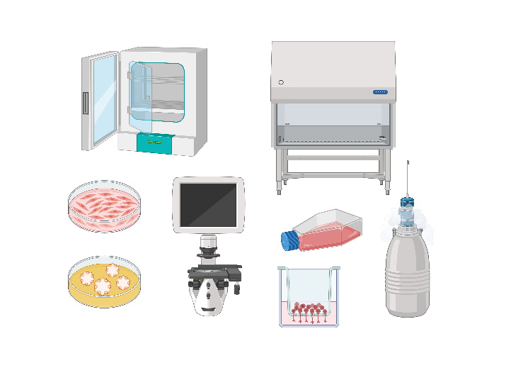
Cell Culture Facility – from conventional cell cultures to advanced ex vivo models
The EPISIGNAL lab’s cell culture facility comprises two fully separated rooms, enabling parallel workflows for standard 2D cultures and advanced 3D systems. One room is dedicated to routine 2D maintenance of immortalized and primary cells, with dedicated biosafety cabinets, CO₂ incubators, and strict SOPs to ensure sterility and reproducibility. The second room supports complex 3D models,
including organoid cultures derived from rodents and patient samples, with dedicated equipment, matrices, and incubation conditions optimized for long-term growth and differentiation. Physical separation and dedicated consumables minimize cross-contamination and allow independent troubleshooting and optimization. The facility supports the full pipeline from sample receipt and processing to expansion, banking, and downstream assays. Routine quality controls—such as mycoplasma screening and viability checks—are embedded in workflows to safeguard data integrity. Ethical handling of human specimens is ensured through documented consent, traceable sample management, and approved protocols. Trained personnel receive ongoing mentoring in aseptic technique, 3D culture handling, and data recording. Together, these features provide a robust platform for modeling disease biology and testing interventions across both simplified 2D systems and physiologically relevant 3D organoids.
Molecular Biology Facility – from genes to proteins, from proteins to function
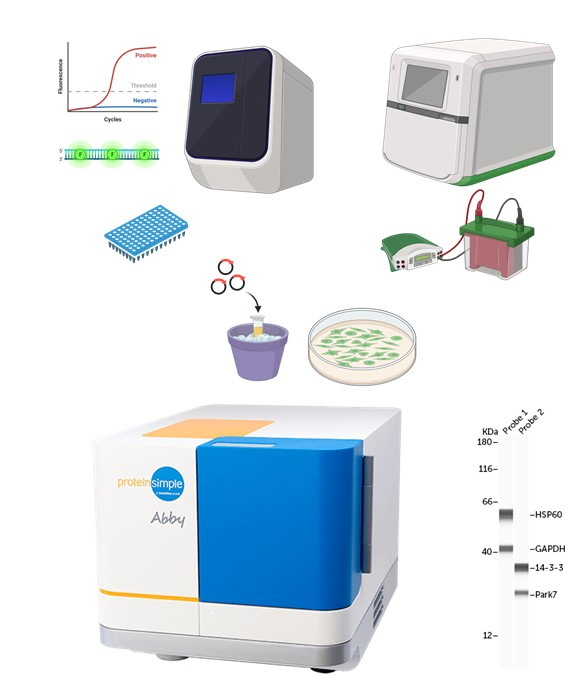
The EPISIGNAL lab’s molecular biology facility supports end-to-end nucleic acid and protein workflows under rigorously maintained SOPs. For transcript analysis, the lab operates real-time qPCR systems enabling both relative (ΔΔCt) and absolute quantification, with validated SYBR Green and probe-based assays and integrated melt-curve quality control. Plate-reader–based assays cover absorbance, fluorescence, and luminescence formats for quantifying nucleic acids, enzymatic activities, viability, and reporter outputs. Standardized
sample prep (RNA/DNA/protein extraction and QC) and calibrated controls ensure reproducible, cross-experiment comparisons. On the protein side, the facility supports conventional Western blotting, from gel casting and transfer to chemiluminescent detection, alongside immunofluorescent stainings for cellular localization on fixed samples. Capillary Western blotting is also available for higher throughput, lower sample input, and improved quantitation, with data exported to common analysis pipelines. Centralized reagent management, training, and documentation round out a platform built for robust, publishable results. The facility features an Abby capillary Western blot system from ProteinSimple. Abby automates the entire Western workflow inside sealed capillaries—separation, immobilization, antibody incubation, detection, and quantitation—reducing hands-on time and variability. It requires low microliter sample volumes and delivers results in hours rather than the typical overnight timeline of traditional blots. The system’s software provides built-in lane (capillary) alignment, background correction, and dynamic range fitting, simplifying objective quantification across runs. By minimizing manual steps and consumables, Abby enables sensitive, reproducible protein expression profiling from limited or precious samples, making it ideal for validation studies and longitudinal projects.